Motor vehicle accident complicated by neurogenic stunned myocardium and neurogenic shock
 Written by Dr. Evan Schmitz, a highly skilled Interventional Pulmonologist and Critical Care Specialist, dedicated to treating the sickest patients with compassionate expertise.
Changes: Initial publication of case study
Abstract
A 25-year-old woman was a restrained driver in a rollover motor vehicle accident (MVA) and suffered a C5-C6 fracture-dislocation with spinal cord injury. She developed neurogenic stunned myocardium and neurogenic shock. Her unopposed parasympathetic nervous system triggered multiple episodes of unstable bradycardia which resulted in multiple cardiac arrests. She was treated with inotropes and vasopressors to keep her mean arterial pressure (MAP) > 85 mmHg for adequate spinal perfusion. Chronotropic medications were used to elevate her heart rate (HR) in order to prevent her unstable bradyarrhythmia from causing a cardiac arrest.
Case presentation
History of present illness:
25-year-old female restrained driver was in a rollover MVA and suffered a C5-C6 fracture with spinal cord injury. (Figure 1).
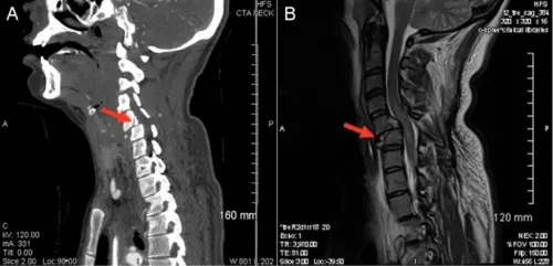
Figure 1. Panel A: computerized tomography (CT) scan of the neck showing C5-C6 fracture and dislocation (arrow). Panel B: Accompanying magnetic resonance imaging (MRI) of the neck. Courtesy: Dr. Schmitz
Physical examination
She was lucid upon evaluation by the trauma team in the emergency room (ER) but was unable to move her lower extremities. She could move her upper extremities and follow commands. Upon arrival to the intensive care unit (ICU) she was tachycardic, tachypneic, hypotensive and hypothermic. Her chest felt full; no crepitus was appreciated.
Hospital course
She was initially resuscitated with 3 liters (L) of fluid and dopamine to maintain her blood pressure (BP). She was externally warmed with a Bair Hugger. It was reported that her HR dropped along with her BP during imaging studies.
Upon initial evaluation in the intensive care unit (ICU) she was found to be in shock while on 15 mcg/kg/min of dopamine and was requiring significant ventilator support with elevated peak and plateau airway pressures. Bedside ultrasound of the left anterior second intercostal space revealed a sliding lung sign, however, a pneumothorax was still suspected. Chest X-ray (CXR) showed a lateral left pneumothorax without any fractured ribs. A left thoracostomy tube was placed with relief of air upon entry into chest cavity. A follow up CXR confirmed resolution of the pneumothorax. The ventilator peak and plateau pressures decreased.
Her BP remained low as well as her HR. Bedside ultrasound of the chest revealed a hypokinetic heart with a small right ventricle and a left ejection fraction (EF) estimated at <40%. No evidence of tamponade. Electrocardiogram (ECG) revealed sinus tachycardia without significant ST changes (Figure 2).

Figure 2. Initial ECG. Courtesy: Dr. Schmitz
Dopamine was discontinued and norepinephrine was started. Another 3 L of fluid were given but the right ventricle remained small upon repeat ultrasound. Her HR remained below 90 beats per minute (bpm) after increasing norepinephrine to 30 mcg/min. While receiving norepinephrine at 50 mcg/min her HR slowly decreased into the 20s and then her pulse was no longer palpable. A code blue was called. Epinephrine and atropine were given and her HR increased and her pulse and BP returned.
A few hours later while on 50 mcg/min norepinephrine, her HR dropped again into the 20s and another code blue was called. An official echocardiogram was performed which confirmed apical/lateral hypokinesia and an EF of approximately 30-35% (Figure 3).

Figure 3. Representative static images from the cardiac ultrasound.
Panel A: systole. Panel B: diastole.1 Courtesy: Dr. Schmitz
She was started on an epinephrine drip. Multiple codes occurred throughout the night with a similar pattern of bradycardia and no pulse. Isoproterenol at 0.016 mcg/kg/hr was started and her HR stayed above 100 bpm. As long as her HR remained in the 120s to 130s her BP was compatible with life.
On day two she was requiring epinephrine at 30 mcg/min, norepinephrine at 50 mcg/min and isoproterenol at 0.016 mcg/kg/hr. Her troponin peaked at 10 ng/mL (upper limit of normal 0.04 ng/mL) and her lactate at 9 mmol/L. By day three remarkably her lactate decreased to 3 ng/mL, her troponin to 6 ng/mL, and her kidney function was normal.
She was alert, following commands and able to move her upper extremities. Propofol and fentanyl were discontinued to avoid further myocardial depression and a ketamine drip with prn midazolam pushes were started. By the afternoon norepinephrine and epinephrine were weaned off and transcutaneous pacer pads were placed with a backup rate of 70 BPM. Phenylephrine was added to the isoproterenol and a MAP > 85 mmHg was maintained to optimally perfuse her spinal cord.
Late in the evening her HR dropped but transcutaneous pacing was able to keep her HR above 70 BPM. Epinephrine was resumed and her HR stabilized in the 130s while on 5 mcg/min of epinephrine and phenylephrine at 420 mcg/min with a MAP > 65 mmHg. She became hyperthermic and she was cooled with an Artic Sun medical device.
Early in the morning on the fourth day her ventilator peak and plateau pressures increased into the 40s and a CXR and bronchoscopy was emergently performed. There was no evidence of pneumothorax or airways obstruction. The CXR revealed multi-lobar patchy infiltrates consistent with adult respiratory distress syndrome (ARDS). Her partial pressure of oxygen (PaO2) ranged from 50 to 70 mmHg until she once again developed bradycardia which did not respond to transcutaneous pacing or atropine. Another code blue was called as her pulse disappeared. After five minutes of cardiopulmonary resuscitation (CPR) return of spontaneous circulation (ROSC) occurred. Once again, her HR and BP began to drop; her family decided to withdraw care.
Discussion
When evaluating a trauma patient after a motor vehicle accident, a high index of suspicion should be maintained for both blunt cardiac injury and a pneumothorax. Blunt chest trauma may cause a myocardial contusion, tamponade as well as a tension pneumothorax. Ultrasound of her heart did not reveal a pericardial effusion or tamponade. Ultrasound of her lungs revealed a sliding lung sign which is fairly sensitive for excluding pneumothorax2, however, in this case the CXR did reveal a left pneumothorax which was decompressed by the placement of a left thoracostomy tube. Despite the resolution of her pneumothorax and resuscitation with 6 L of fluid there was no improvement in her hypotension. In the absence of hemorrhage, this makes intravascular volume depletion unlikely the cause of her hypotension.
She was diagnosed with cardiogenic shock from neurogenic stunned myocardium (NSM). NSM has been described in subarachnoid hemorrhage, intracerebral hemorrhage, acute ischemic stroke and head trauma.3 It is thought to be similar to stress-induced cardiomyopathy (Takatsubo’s cardiomyopathy) or apical ballooning syndrome. NSM is thought to be initiated by an injury to the hypothalamus. This causes a surge in adrenergic output with a resultant increase in norepinephrine release within the myocardium. Plasma concentrations remain in the normal range. This surge leads to myocyte calcium overload and contraction band myonecrosis with resultant systolic heart failure. Studies using myocardial scintigraphy have confirmed normal perfusion with normal MIBI (Technetium-99m Sestamibi) uptake with abnormal MIBG (metaiodobenzylguanidine) uptake correlating with the regional wall motion abnormalities seen in patients who develop NSM. The resultant functional myocardial sympathetic denervation leads to cardiac injury and release of troponin. Supportive care consists of inotropes and vasopressors. The goal is to minimize these medications, if possible, to allow for healing of the myocardium which usually occurs within a few weeks.
Although neurogenic stunned myocardium explains the patient’s systolic heart failure, it does not explain her diminished preload after fluid resuscitation and her symptomatic bradycardia with persistent shock. Neurogenic shock caused by her spinal cord trauma resulted in a loss of sympathetic tone but retention of her parasympathetic tone. The lack of sympathetic tone explains her diminished pre-load because it caused her vasculature to dilate and decrease the venous return to the heart. Retention of her parasympathetic tone caused her to become bradycardic.
Neurogenic shock is caused by traumatic sympathetic nerve injury to the spine above T6 level.4 This leads to a lack of sympathetic tone and unopposed parasympathetic response. The dysregulation of the autonomous nervous system causes organ tissue hypoperfusion by vasogenic dilation of the blood vessels. Patients are at risk of unopposed vagal stimulation which can cause severe bradycardia and may lead to cardiac arrest as seen in this patient. The treatment is vasopressor support with a mean arterial pressure (MAP) goal up 85-90 mmHg to ensure adequate spinal perfusion. Chronotropic medications such as atropine, an acetylcholine receptor antagonist as well as isoproterenol, a beta-adrenergic agonist, are used to treat bradycardia and prevent pulseless electrical activity (PEA) cardiac arrest.
The reason her HR responded to epinephrine and isoproterenol is because both of these medications cause an increase in sinoatrial node (SA) firing and conduction through the atrioventricular (AV) node by beta-adrenergic stimulation which opposes the cholinergic effects of the parasympathetic system. In addition, alpha-adrenergic stimulation from epinephrine causes vasoconstriction.
Her BP responded well to the addition of phenylephrine, an alpha-adrenergic agonist, when her epinephrine was weaned off and she was able to maintain a MAP >85 mmHg until she developed ARDS.
Once she developed ARDS her oxygen delivery became severely compromised. This caused her body to rely more on anaerobic metabolism which gave rise to the production of more lactic acid. Her lungs and kidneys were unable to compensate as her metabolic acidosis worsened. As her PaO2 decreased and her lactic acid increased her heart failure worsened which resulted in cardiac arrest.
Conclusion
Motor vehicle accidents can cause severe traumatic injuries. It is important to be aware of all the cardiac complications that may arise from these traumatic events and treat those conditions appropriately. Traumatic head injuries can cause neurogenic stunned myocardium which can lead to cardiogenic shock. Spinal trauma can cause neurogenic shock by dilating blood vessel and limiting pre-load and may also cause unstable bradycardia. These devastating complications of MVA are managed with inotropes, vasopressors, and chronotropic medications.
References
- Schmitz ED. Ultrasound for critical care physicians: hypotension after a MVA. Southwest J Pulm Crit Care. 2014;8(3):176-8. doi: https://www.swjpcc.com/critical-care/2014/3/4/ultrasound-for-critical-care-physicians-hypotension-after-a.html
- Husain LF,Hagopian L,Wayman D, BakerWE, CarmodyKA. Sonographic diagnosis of pneumothorax. J Emerg Trauma Shock. 2012;5(1):76–81
- Banki NM, Kopelnik A, Dae MW, et al. Acute neurocardiogenic injury after subarachnoid hemorrhage. Circulation 2005;112(21):3314-9.
- Dave, S, Dahlstrom, J; Weisbrod L. Neurogenic shock. StatPearls 2025. https://www.ncbi.nlm.nih.gov/books/NBK459361/
Test your knowledge
Stay on track!
Would you like a reminder when your ACLS certification expires, plus study tips?
How we reviewed this article
Our experts continually monitor the medical science space, and we update our articles when new information becomes available.
- Current versionMail the author of this pageEmail
- Oct 22, 2025
Written by:
Changes: Initial publication of case study